Art of Science 2022 finalists

Art of Science 2022 finalists

Discover the beauty and wonder of Art of Science

What would you like to do?


Still images


Against the grain by Kristy Shield-Artin and Suzan Sam 

Blue cancer cells swirl around a yellow centre of secreted mucin in a tumour of the lung from a patient who has developed and survived three different cancers over more than a decade. The surrounding lung tissue in green shows reactive changes. 


SECOND PLACE - STILL IMAGE CATEGORY

At ease by Bianca Capaldo 

Breast tissue is seen in its resting state - luminal cells (purple) within an outer layer of myoepithelial cells (green). During breastfeeding, luminal cells grow and change to produce milk, which the myoepithelial cells propel along the ducts. 


Chemokine chrysalis by Raymond Qin 

Within a lymph node, small proteins called chemokines guide immune cells to distinct locations in response to foreign antigens, including cancer cells and infectious agents. The different colours in the image identify cells that produce different chemokines.


Art of Science image

Crypto rain by Stefanie Bader      

The sparkling ‘raindrops’ in this image are Cryptosporidium parasites, which cause a water-borne diarrhoeal illness responsible for 200,000 deaths per year around the world. Research is investigating different parasite components as potential targets for vaccines. 


Art of Science image

In bloom by Caleb Dawson 

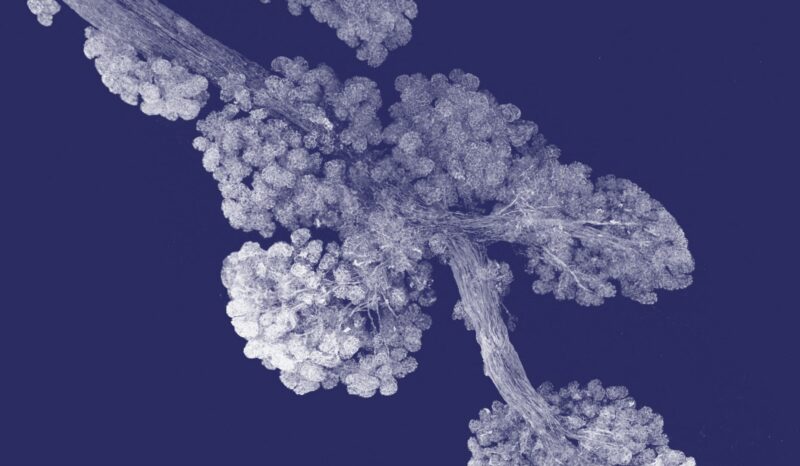
In response to hormones released during lactation, the ducts and lobules of breast tissue ‘bloom’ to form clusters of alveoli. These contain milk-producing cells encircled by tiny muscle-like cells, which contract to deliver milk to an infant. 


Art of Science image

FIRST PLACE - STILL IMAGE CATEGORY

Interweb of the mind by Melody Leong  

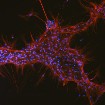
This image shows two different types of nerve cells or neurons (depicted in red and blue) grown in the laboratory. Researchers are using these ‘cultured’ neurons to study how genetic mutations contribute to neurodevelopmental disorders associated with intellectual disability. 


Art of Science image

Keep an eye out by Melanie Dietrich 

These 3D X-ray images show microscopic crystals formed by malaria parasite proteins interacting with ‘nanobodies’, small antibodies that can block parasite development and transmission.


Art of Science image

Light in the dark by Kristy Shield-Artin and Suzan Sam 

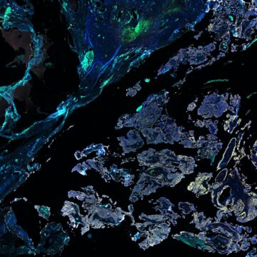
In this image of an ovarian tumour, different cell and tissue types have been stained with multiple fluorescent labels. Cyan specks identify immune cells whose role in tumour growth and response to treatment are under investigation.


Art of Science image

Supernova by Stefanie Bader and Kathryn Davidson     

This image shows organoids - miniature laboratory models of organs grown from patient biopsies - being used to test drugs targeting infections of the liver. The top ‘exploding’ liver organoid was treated with a drug that triggers the death of infected cells.


Art of Science image

THIRD PLACE - STILL IMAGE CATEGORY

Through the looking glass by Sabrina Lewis 

Each colour in this image represents a unique cancer cell type within a breast tumour. Researchers are studying how different cancer cells spread to distant organs to form metastases, which can have dire consequences for patient survival. 


Art of Science image

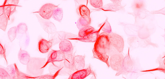
Toxo Actin’ Chill by Aurelie Dawson, Caleb Dawson and Cindy Evelyn 

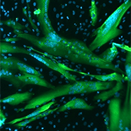
Toxoplasma gondii  (‘Toxo’) parasites responsible for the infectious disease toxoplasmosis are shown ‘hiding’ inside a host cell to evade detection by the immune system. Toxo are pictured in yellow; the red filaments are the support ‘skeletons’ of the host cells. 


Return to top of page


Moving images

Watch our full Art of Science 2022 video playlist on YouTube.


Bad aberrations by Michael Mlodzianoski 

Even high-tech modern microscopes are subject to aberrations that reduce the resolution and accuracy of images. The video shows the effects of different aberrations on the point spread function (PSF), a measure of image quality.


Red forest by Sabrina Lewis             

This animation takes us on a 3D ride around and through the forest of blood vessels in a section of the lung. Research is examining how tumour cells in the lungs interact with blood vessels.


Popcorn Toxo by Aurelie Dawson and Caleb Dawson 

The common and usually harmless parasite Toxoplasma gondii can cause the serious disease toxoplasmosis in infants born to infected mothers and in people with weakened immune systems. The video shows ‘Toxo’ parasites multiplying and flashing brightly as they burst out of infected cells.


THIRD PLACE - MOVING IMAGE CATEGORY

The tree of life by Kelin Zhao         

This video shows a 3D view of the tree-like network of blood vessels in the thymus. Through this, precursors of immune cells called T-lymphocytes enter the gland to be selected and trained to recognise and target foreign antigens.


FIRST PLACE - MOVING IMAGE CATEGORY

Thymic coral reef by Kelin Zhao 

The thymus is a vital immune gland where white blood cells called T-cells are ‘trained’ to respond to foreign antigens. The video shows a 3D reconstruction of a thymic lobe: the inner medulla has a large, branched central structure (purple) with small outer islets (various colours). 


Transmission by Niall Geoghegan, Robyn McConville and Justin Boddey 

Only male sexual stage Plasmodium parasites can transmit malaria from humans back to mosquitoes. Here they are seen dividing and bursting out of human red blood cells in the gut of a mosquito, ready to fertilise female parasites. 


Web of lyse by George Ashdown and Niall Geoghegan  

White blood cells called neutrophils are shown ‘weaponizing’ their own DNA - stained fluorescent orange - to trap and destroy pathogens by forming neutrophil extracellular traps (NETs). 


What’s that, neuron? by Melody Leong and Yuqing Yang  

A compilation of high-resolution microscope images shows successive cross-sections throughout a laboratory-grown nerve cell (neuron), which harbours a genetic mutation linked to neurodevelopmental disorders associated with intellectual disability.


SECOND PLACE - MOVING IMAGE CATEGORY

The (not so micro)glia by Matthias Mulazzani  

Microglia - shown in yellow associated with blood vessels in blue - are immune cells resident in the brain. They support nerve cells, playing an important role in brain development and possibly in diseases like dementia, stroke and brain tumours. 


Return to top of page


More information

 
Lattice light sheet microscope

Optical microscopy has become one of the most powerful tools in medical research.

WEHI's Centre for Dynamic Imaging is advancing our understanding of how diseases develop, spread and respond to treatment.

Researcher facing news media crews

Catch up on our latest research discoveries and announcements.

Two researchers smiling at the camera

Want to be informed of our most exciting discoveries? Subscribe to our quarterly newsletter, Illuminate.

Two scientists chatting on a couch

Meet the superstars of science and the talented young sparks of the future.

Hear from the patients whose lives have been changed by new discoveries and find out how investing in WEHI’s brightest minds will help solve our most complex medical challenges.