Institute researchers have uncovered new insights into how the normal controls on cell growth are lost in cancer cells, leading to rapid tumour expansion.
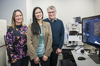
(L-R) Dr Kelly Rogers, Dr Kim Pham and Professor Phil
Hodgkin have uncovered new insights into cancer growth.
The findings could help researchers predict how cancer cells respond to chemotherapy and improve our understanding of how cancer evolves.
Researchers have discovered how the process of cell replication is derailed in cancer cells, challenging a longstanding theory about how cells grow.
The discovery was made using advanced imaging techniques and allowed researchers to develop a new mathematical model of cell replication.
Accurate models of cell replication help researchers predict how cancers respond to chemotherapy and how drug resistance evolves.
Debunking cell cycle dogma
Cell replication is a normal process that generates additional cells, enabling the body to grow, repair tissues and generate germ-fighting immune cells.
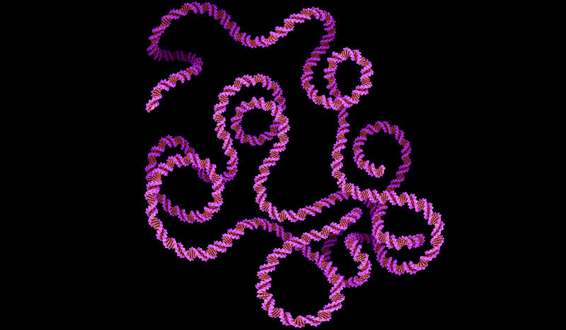
In order to replicate, a cell first copies its genetic material, DNA, and then physically splits in half to form two new ‘daughter’ cells. This process, called the cell cycle, is normally tightly controlled to prevent excessive growth, which can lead to cancer.
In this study, researchers uncovered precisely how the cell cycle is derailed in cancer cells.
“We found that compared with healthy immune cells, cancer cells had dramatic changes in their cell cycle,” Dr Pham said.
“The first phase of the cell cycle, called G1, is normally tightly controlled to ensure replication occurs safely. This step is drastically shortened in cancer cells, allowing them to race through the cell cycle at a risky pace.”
Since 1973, scientists have assumed that the second phase of the cell cycle, when DNA is copied and the cell splits in half, takes a fixed amount of time, while the first phase takes a variable amount of time.
Several years ago, the team challenged this theory in healthy immune cells, showing that both phases of the cell cycle contribute to changes in replication time. Now, they have upended the theory in cancer cells too.
“The old theory predicts that all variation in replication time comes from the first phase of the cell cycle,” Dr Pham said.
“When we looked closely at the cancer cells, we found that the opposite was true: the bulk of the variation was due to the second phase of the cell cycle.”
Imaging cells in unprecedented detail
The discovery was made in collaboration with the Centre for Dynamic Imaging, a laboratory within the Institute that offers researchers access to advanced imaging technologies.
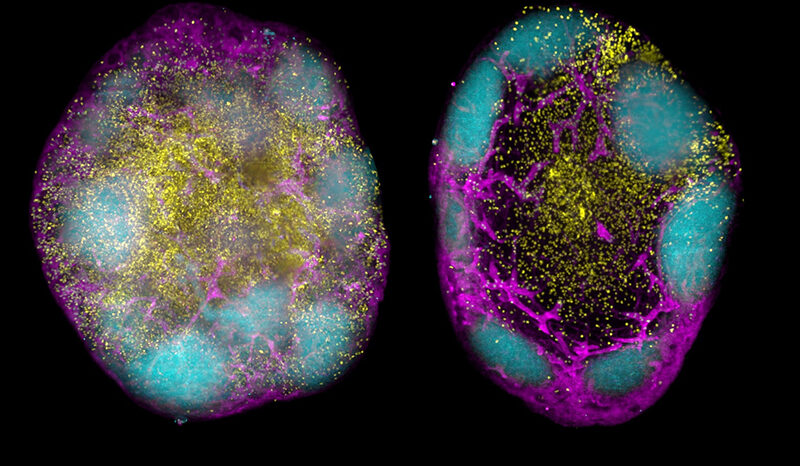
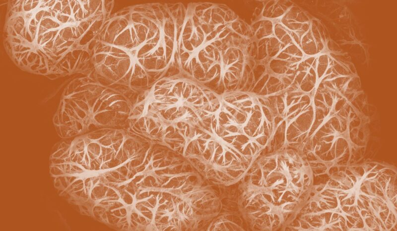
“For this study, we tagged cancer cells with a fluorescent sensor that changes colour as cells progress through the cell cycle,” Dr Pham said. “We then performed single cell imaging to track each phase of the cell cycle as they underwent replication,” she said.
The researchers then worked with bioimage analyst Dr Whitehead to analyse and interpret their data and develop a new mathematical model for predicting when cells replicate.
“This study demonstrates the power of imaging to directly reveal cellular behaviours, and in some cases challenge assumptions that were made before it was possible to obtain such clear evidence,” Dr Whitehead said.
Fresh insights into cancer
Professor Hodgkin said the research could impact on our understanding of cancer.
“Accurate mathematical models of how cancer cells replicate help us predict how cancers respond to chemotherapy treatment, and how they evolve to become drug resistant.”
The team’s finding that the first phase of the cell cycle is minimised in cancer cells suggests a vulnerability that could be targeted by cancer treatments.
“Cancers have often lost the safety checks that prevent replication in the presence of errors such as DNA damage,” Professor Hodgkin said. “Our work suggests the lack of these safety checks leads to the first phase of the cell cycle becoming much shorter in cancer cells.
“Drugs that help restore these safety checks could be beneficial for treating multiple cancers.”
The research was supported by the National Health and Medical Research Council, Human Frontiers Science Program and the Australian Government.