WEHI researchers have developed enhanced imaging technology that can model how breast cancer cells invade and spread into bone and remodify themselves to fuel tumour growth.
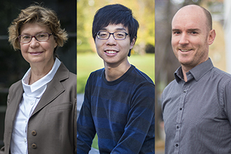
Pictured: Professor Jane Visvader, Dr Raymond Yip and Associate Professor Edwin Hawkins.
The team found that tumour spread occurred at specific locations in the bone, not randomly as previously thought. They also showed that breast cancer cells ‘renovate’ the bone to create an environment that fuels their spread, while starving the body of essential nutrients.
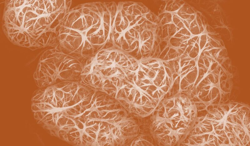
Using WEHI’s Centre for Dynamic Imaging, the research team took hundreds of images to create three-dimensional (3D) models of the bone marrow and the blood vessels that run through them – some as tiny as a fraction of a micron. These images were used to better understand why breast cancer cells metastasise (spread) into bone, and what factors facilitate their growth.
Researchers developed cutting-edge 3D technology to map how breast cancer cells invade and spread into secondary sites in the bone.
They found tumour spread occurred at specific locations in the bone, where breast cancer cells ‘remodel’ the bone to create an environment that fuels its growth.
The research could help to guide development of new therapies for patients suffering from cancers that typically spread to the bone, including breast cancers and prostate cancers.
Radical renovation
Bone marrow is one of the most common sites of metastasis in people with breast cancer. Spread of cancers to secondary organs, such as the bone, is often incurable, and breast cancer patients with bone metastases typically have a very poor prognosis.
Dr Yip said the research provided a fascinating insight into how cancer cells colonise the bone marrow, by mapping out the vessels and the tumour structures next to them.
“We think of bones as these static, structural organs – but they’re highly dynamic”, he said.
“Our research shows breast cancer cells preferentially reside near a specific blood vessel subtype in the bone called the type H vessels. In other words, breast tumour cells selectively home to a specialised vasculature, suggesting type H vessels are supplying certain growth factors to nurture breast cancer cells growth in bone.”
Associate Professor Hawkins said the idea that cancer cells can remodel their environment isn’t new, however it hadn’t been explored in a “tricky organ” like bone marrow until now.
“What Raymond showed us through his incredible 3D images is that a tumour cell will move to the bone marrow and completely renovate that home” Associate Professor Hawkins said.
“We saw that when breast cancer cells metastasise to the bone marrow, they release tumour-derived growth factors that enable them to ‘remodel’ to create a favourable environment that further facilitates their growth – unfortunately at the detriment of the whole body.”
By delving into the symbiotic relationship between our host bodies and these tumour cells, the researchers hope their discoveries can one day lead to a better understanding of mechanisms that these cells work with that can make them easier to treat or prevent tumour spread.
Cancer treatment enhancements
While breast cancer patients are the focus of the current research, spread of tumours to the bone is common in other cancers, including prostate, lung, kidney and thyroid cancers and melanoma.
Professor Visvader said these mechanisms could be a target for future therapeutic discovery.
“Cancer therapeutic discovery has expanded considerably over past decades to not only target the cancer cells directly, but also the mechanisms used by cancer cells to enhance their growth,” she said.
“It will be important to further understand the mechanisms by which tumour cell-derived factors remodel blood vessels, as this could help define new therapies for patients in the future. We also hope to take this innovative 3D imaging technique we have developed and extend it to other diseases that involve bone metastases.”
The research was supported by the Australian Cancer Research Foundation, the Australian National Health and Medical Research Council, and the Victorian Government.
WEHI authors
Raymond Yip, Joel Rimes, Bianca Capaldo, François Vaillant, Kellie Mouchmore, Bhupinder Pal, Yunshun Chen, Elliot Surgenor, Gordon Smyth, Geoff Lindeman, Edwin Hawkins, Jane Visvader.