Flow cytometry

Flow cytometry

Flow cytometer instrument
Flow cytometry is a versatile technology used to study the composition of mixtures of cells. Analysers reveal this composition and cell sorters can physically separate cells for further study.

Flow cytometers can be used to identify and separate:

  • Different types of cells
  • Cells at different stages of their life cycle
  • Activated cells
  • Dying cells
  • Cancerous cells
  • Chromosomes
  • Microorganisms

Flow cytometry is used in:

  • Hospitals, for diagnosis and treatment
  • Medical research laboratories
  • Marine biology
  • Microbiology
  • Agricultural industries

Flow cytometry at the Institute

The first flow cytometer in the southern hemisphere, the FACS II, was introduced here at the Institute in 1977. Since then flow cytometry technology has continued to advance and is now a key resource for researchers.

Flow cytometry is widely used at the Institute where researchers often need to profile samples containing mixtures of cells. For example:

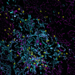
  • Immunology researchers use flow cytometry to identify, separate and define various immune cell subtypes by their size and structural characteristics.
  • Researchers using CRISPR/Cas9 genome editing technology tag edited genes with fluorescent proteins, allowing cells to be detected and separated by flow cytometry.

The Institute’s flow cytometry resources are also available for external use. 

How does flow cytometry work?

In a flow cytometer:

  • Cells are transported in a fluid stream into the path of one or more focussed laser beams.
  • Cells containing fluorescent dyes absorb light, which is then re-emitted at longer wavelengths.
  • Sensors detect and measure the many different wavelengths of light from each cell.
  • The resulting data file describes the composition of the mixture of cells.

Fluorescent tags can be used to identify various proteins both within and on the surface of cells as well as tags that bind DNA, RNA and various other cellular components.

About the animation: cells are delivered into the path of a focused blue laser beam. Cells with no label scatter some blue light. Cells labelled with a yellow or red fluorescent dye give off either of yellow or red pulse of light as well as scattered blue light. The scattered and fluorescent light pulses from many thousands of cells are measured and can be represented graphically to describe the composition of the sample.

Researchers: 
Add This: